
| Prostasin (Human) ELISA Kit | |
| A Biomarker for Carcer and Preeclampsia Research | |
Increased expression of prostasin contributes to early-onset severe preeclampsia through inhibiting trophoblast invasion |
|
| Objective:To investigate the potential role of prostasin, as an invasion suppressor, in the process of trophoblast invasion in preeclampsia.Study design:This case-control study included 19 early-onset severe preeclampsia (⩽34 weeks), 20 late-onset severe preeclampsia (>34 weeks) and 20 normal term pregnant women. Immunohistochemistry was conducted to identify the cellular localization of prostasin, as well as the matrix metalloproteinase 2 (MMP2) and MMP9 in the placenta tissues. Enzyme-linked immunosorbent assay was performed to analyze the expression of these three proteins in placental homogenates. The effect of prostasin on the invasive and migratory ability of trophoblast cells was detected by transwell assays. We also examined the regulation of the prostasin antibody in the MMP2 and MMP9 secretion by HTR-8/SVneo cells via blocking theprostasin activity.Result:This study demonstrated that the prostasin, MMP2 and MMP9 were all expressed in the placental syncytiotrophoblasts. Increased expression of prostasin was detected in cases with early-onset severe preeclampsia compared with the late-onset and control groups (P<0.05), whereas the expression patterns of MMP2 and MMP9 in placental homogenates were opposite to that of prostasin (P<0.05). Recombinantprostasin inhibited the invasion and migration of trophoblast cells, whereas prostasin antibody enhanced the MMP2 and MMP9 secretion in a dose- and time-dependent manner.Conclusion:These findings suggest that prostasin may suppress the invasion process in preeclampsia by attenuating MMP2 and MMP9 secretion | |
| Yang Y et al. J Perinatol. 2014 Jul 31. doi: 10.1038/jp.2014.136. [Epub ahead of print] | |
Axl and prostasin are biomarkers for prognosis of ovarian adenocarcinoma |
|
| In this study, the protein levels of Axl and prostasin in malignant neoplasms of the ovary and their clinicopathologic significance were investigated. The protein levels of Axl and prostasin in ovarian adenocarcinomas (n = 80), serous cystadenoma (n = 15), mucinous cystadenomas (n = 15), and normal ovary tissues (n = 10) were measured using immunohistochemistry. The percentage of Axl-positive cases was significantly higher in ovarian adenocarcinoma (61.3%) than in mucinous adenoma tissues (13.3%; P < .001) and normal tissues (0.0%; P = .000). The percentage of prostasin-positive cases was significantly lower in ovarian adenocarcinoma (42.5%) than in mucinous adenoma tissues (86.7%; P = .000) and normal tissues (100%; P = .000). The expression of Axl was significantly lower in cases with G1 tumor and TNM stage I or II tumor with no lymph node metastasis than in cases with G3 tumor and TNM stage III or IV tumor with lymph node metastasis (P < .05 or P < .01). However, the expression pattern of prostasin was opposite to that of Axl (P < .01 or P < .01). Univariate Kaplan-Meier analysis showed a negative correlation between Axl expression (P = .000) and overall survival and a positive correlation between prostasin expression (P = .000) and overall survival. Multivariate Cox regression analysis showed that Axl-positive expression and prostasin-negative expression are independent bad prognostic predictors in ovarian adenocarcinoma. Our study suggested that Axl and prostasin expression may be closely related to carcinogenesis, metastasis, and prognosis of ovarian adenocarcinoma. Ann Diagn Pathol. 2013 Oct;17(5):425-9. doi: 10.1016/j.anndiagpath.2013.01.005. Epub 2013 May 23. |
|
Comprehensive serum profiling for the discovery of epithelial ovarian cancer biomarkers |
|
| FDA-cleared ovarian cancer biomarkers are limited to CA-125 and HE4 for monitoring and recurrence and OVA1, a multivariate panel consisting of CA-125 and four additional biomarkers, for referring patients to a specialist. Due to relatively poor performance of these tests, more accurate and broadly applicable biomarkers are needed. We evaluated the dysregulation of 259 candidate cancer markers in serum samples from 499 patients. Sera were collected prospectively at 11 monitored sites under a single well-defined protocol. All stages of ovarian cancer and common benign gynecological conditions were represented. To ensure consistency and comparability of biomarker comparisons, all measurements were performed on a single platform, at a single site, using a panel of rigorously calibrated, qualified, high-throughput, multiplexed immunoassays and all analyses were conducted using the same software. Each marker was evaluated independently for its ability to differentiate ovarian cancer from benign conditions. A total of 175 markers were dysregulated in the cancer samples. HE4 (AUC=0.933) and CA-125 (AUC=0.907) were the most informative biomarkers, followed by IL-2 receptor a, a1-antitrypsin, C-reactive protein, YKL-40, cellular fibronectin, CA-72-4 and prostasin (AUC>0.800). To improve the discrimination between cancer and benign conditions, a simple multivariate combination of markers was explored using logistic regression. When combined into a single panel, the nine most informative individual biomarkers yielded an AUC value of 0.950, significantly higher than obtained when combining the markers in the OVA1 panel (AUC 0.912). Additionally, at a threshold sensitivity of 90%, the combination of the top 9 markers gave 88.9% specificity compared to 63.4% specificity for the OVA1 markers. Although a blinded validation study has not yet been performed, these results indicate that alternative biomarker combinations might lead to significant improvements in the detection of ovarian cancer. | |
| Yip P, et al. PLoS One. 2011;6(12):e29533. doi: 10.1371/journal.pone.0029533. Epub 2011 Dec 21 | |
 |
|
| HumanProstasin ELISA Kit Code No.: SK00156-06 Size: 96T Price: $420.00 USD Standard Range: 39- 2500 pg/mL Sensitivity: 10 pg/mL Sample Type: serum, EDTA Plasma Sample volume: 100 uL Dilution factor: Optimal dilutions should be determined by each laboratory for each application IntraCV: 4-6% InterCV: 8-12% Protocol: PDF |
|
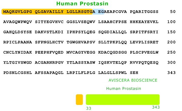
| Human Prostasin Recombinant Code No.: 00156-01-100 Size: 100 ug Price: $360.00 USD Protein ID:NP_002764 Gene ID:NM_002773 MW:50.34 KDa Tag: His Tag on N-Terminus Expressed: E. Coli Purity: 95% Data Sheet: PDF |
|
| Anti Human
Prostasin IgG Code No.: A00156-01-100 Size: 100 ug Price: $260.00 USD Host: Rabbit Antigen: mature form of Prostasin (Human) Ab Type: Polyclonal IgG Purification: Protein A Applications: E,IHC Working Dilution: E: 0.125 ug/ml ; IHC 2-4 ug/ml Data Sheet: PDF |
|
|
|
| Name | Code No. |
Size |
Price ($) |
| Prostasin(Human) ELISA Kit | SK00156-06 | 96 T | 420.00 |
| control for Prostasin (Human) ELISA Kit |
156-06-04 |
each |
120.00 |
| Standard for Prostasin (Human) ELISA Kit | 156-06-02 |
each |
140.00 |
| Detection Antibody for Prostasin (Human) ELISA Kit | 156-06-03 |
96 T |
140.00 |
| Capture Plate for Prostasin (Human) ELISA Kit | 156-06-01 |
96 T |
140.00 |
| Prostasin (Human) rec | 100 ug |
360.00 |
|
| Prostasin (Human) rec | 00156-01-50 |
50 ug |
250.00 |
| Anti Prostasin (Human) IgG | 100 ug |
260.00 |
|
| Anti Prostasin (Human) IgG | A00156-01-50B |
50 ug |
360.00 |